ELECTRICAL SYNAPSE
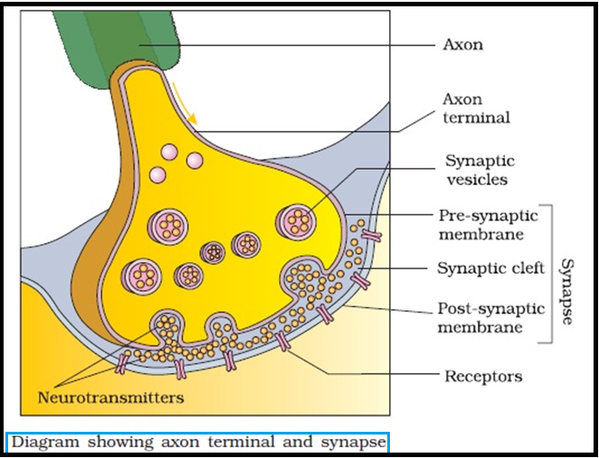
● A `color{violet}("nerve impulse")` is `color{violet}("transmitted")` from one `color{violet}("neuron")` to another through junctions called `color{brown}("synapses.")`
● A `color{brown}("synapse")` is formed by the membranes of a `color{violet}("pre-synaptic neuron")` and a `color{violet}("post-synaptic neuron")`, which may or may not be separated by a gap called `color{brown}("synaptic cleft.")`
● There are two types of `color{violet}("synapses,")` namely, `color{brown}("electrical synapses")` and `color{brown}("chemical synapses.")`
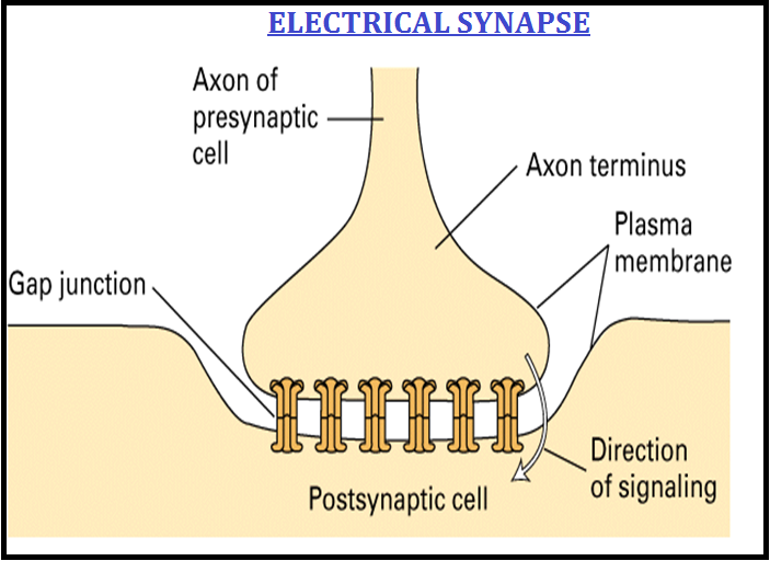
● At `color{violet}("electrical synapses,")` the membranes of `color{violet}("pre- and post-synaptic")` neurons are in very `color{brown}("close proximity.")`
● `color{violet}("Electrical current")` can flow directly from one `color{violet}("neuron")` into the other across these `color{violet}("synapses.")`
● `color{violet}("Transmission of an impulse")` across `color{violet}("electrical synapses")` is very similar to impulse conduction along a `color{violet}("single axon.")`
● `color{violet}("Impulse transmission")` across an `color{violet}("electrical synapse")` is always `color{brown}("faster")` than that across a `color{violet}("chemical synapse.")`
● `color{violet}("Electrical synapses")` are rare in our system
● A `color{brown}("synapse")` is formed by the membranes of a `color{violet}("pre-synaptic neuron")` and a `color{violet}("post-synaptic neuron")`, which may or may not be separated by a gap called `color{brown}("synaptic cleft.")`
● There are two types of `color{violet}("synapses,")` namely, `color{brown}("electrical synapses")` and `color{brown}("chemical synapses.")`
● At `color{violet}("electrical synapses,")` the membranes of `color{violet}("pre- and post-synaptic")` neurons are in very `color{brown}("close proximity.")`
● `color{violet}("Electrical current")` can flow directly from one `color{violet}("neuron")` into the other across these `color{violet}("synapses.")`
● `color{violet}("Transmission of an impulse")` across `color{violet}("electrical synapses")` is very similar to impulse conduction along a `color{violet}("single axon.")`
● `color{violet}("Impulse transmission")` across an `color{violet}("electrical synapse")` is always `color{brown}("faster")` than that across a `color{violet}("chemical synapse.")`
● `color{violet}("Electrical synapses")` are rare in our system